Saturday, March 30, 2013

Article Review (1996) - A Frame Shift Mutation in the Fibrinogen A Alpha Chain Gene in a Kindred With Renal Amyloidosis

We previously learned that the first published report of a fibrinogen mutation that causes familial amyloidosis occurred in 1993 with the Arg554Leu mutation, followed in 1994 with the reporting of the Glu526Val mutation. This post reviews the article describing the next mutation that was reported, two years later in 1996. There are a couple of significant differences between this mutation and the two previously reported mutations. One is the type of mutation itself, and the other is regarding the symptoms.

Title: A Frame Shift Mutation in the Fibrinogen A Alpha Chain Gene in a Kindred With Renal Amyloidosis (1)

Authors: Tomoyuki Uemichi, Juris J. Liepnieks, Toshiyuki Yamada, Morie A. Gertz, Nils Bang, and Merrill D. Benson (Indiana University School of Medicine, Indianapolis, IN, USA and Mayo Clinic, Rochester, MN, USA)

Journal: Blood (1996)

Abstract:
A new American kindred with amyloidosis was found by single-strand conformation polymorphism analysis to have a mutation in the fibrinogen A alpha chain gene. Affected members in this kindred have autosomal dominant amyloid nephropathy. DNA sequencing showed a single nucleotide deletion at the third base of codon 524 of the fibrinogen A alpha chain genes (4904delG) that resulted in a frame shift and premature termination of the protein at codon 548. Antiserum was produced to a portion of the abnormal peptide predicted by the DNA sequence and amyloid deposits were immuno-histologically proven to contain this abnormal peptide. Two of the propositus' 4 children were positive for the mutant fibrinogen A alpha chain gene by restriction fragment length polymorphism analysis based on polymerase chain reaction. These two mutant gene carriers now in the second decade of life show no clinical symptoms of amyloidosis as yet but have lower plasma fibrinogen concentrations when compared with their normal siblings. This [is] the first description of a kindred with renal amyloidosis and low plasma fibrinogen and also the first report of amyloidosis caused by a frame shift mutation.
Here is a link to the PDF of the article, if you'd like to follow along: http://bloodjournal.hematologylibrary.org/content/87/10/4197.full.pdf

As mentioned in the abstract, this article reports on a US kindred found to have a fibrinogen mutation. The propositus (first family member diagnosed) was a woman who was diagnosed with proteinuria at the age of 41 and died after developing cardiac failure and renal insufficiency at the age of 46. A kidney biopsy at the age of 42 showed amyloid deposits in the glomeruli. An autopsy showed amyloid deposits in both kidneys but not in the heart.

Her mother died at the age of 38 due to renal amyloidosis, and one of her mother's brothers died of renal failure at the age of 41.

We'll skip the Materials and Methods section since it's just the details of the laboratory processes of isolating fibrinogen from the affected patient, autopsied tissues, and possibly affected relatives.

The Results section of the article describes the actual mutation that was discovered. This mutation was on the fibrinogen A alpha chain, but instead of a substitution like the previous two mutations (where a G, A, T or C at one position in the DNA is substituted for one of the other three letters), this mutation was a deletion at one position (4904), which shifted everything else beyond that position by one. (That's why they use the term "frame shift mutation.") Since these letters are in groups of three to determine each amino acid in the sequence, each amino acid after this point was also wrong because of this shift. But not only that, this shift also caused the string to end prematurely 25 amino acids later because a particular three-letter sequence came up that signals the end of the string. I'll give a musical analogy to help you picture what this frame shift mutation really means.

Think of the four main building blocks of DNA (represented by the letters G, A, T and C) as musical notes. These four building blocks assemble in groups of three to form amino acids, so think of amino acids as three-note musical chords. In the case of a substitution mutation such as Glu526Val, when these chords are played in order everything sounds fine up to the point of the mutation, then there is one wrong chord, then everything sounds fine after that. But in the case of a frame shift mutation like the one in this article, all the chords sound fine until the mutation is reached, and then every chord after that is likely to be wrong because it will have two notes from the correct chord and one note from the next chord. Plus, in this particular case, the chords stop prematurely.

Next they describe the other interesting difference between this mutation and the previously described mutations. You may recall that the 1994 article showed the Glu526Val mutation had no impact on blood clotting times. The mutation described in this article from 1996, however, does seem to have an impact on blood clotting times and plasma fibrinogen concentration levels. They found the blood clotting times were prolonged, and plasma fibrinogen concentration levels were at or below normal levels. These changes were not enough to be classified as hypofibrinogenemia (a deficiency of fibrinogen in the blood), but there is some measurable impact. So if you have this particular mutation you should inform your doctor that there is an impact on blood clotting times. (I suppose that should be considered when prescribing anticoagulants, for instance.)

The Discussion section of the article begins with a general overview of the structure of fibrinogen and various mutations known to cause blood clotting disorders. It then states there are three kindreds in previously published reports that have fibrinogen A alpha chain mutations known to cause hereditary renal amyloidosis. So the kindred in this report is only the fourth one to be reported with such a mutation. (Three mutations, four families.)

Although they could not test any tissue samples from the propositus' mother, it is likely that she had the mutation because she died of renal amyloidosis at the age of 38, and the propositus' father does not have the mutation. (The Results section also mentioned that two of the propositus' four children tested positive for the mutation but do not have any symptoms.)

The age of onset seems to be different for the three mutations that have been reported up to this point. The earliest age of onset occurs with the first reported mutation (Arg554Leu). Those patients were affected in their 20s or 30s. For the frameshift mutation in this article, the age of onset appears to be in the late 30s or early 40s. Finally, for the Glu526Val mutation, the age of onset has been in the 40s through 60s.

This section then has a paragraph about treatment. Hemodialysis is mentioned since that is a standard treatment for other renal diseases. Regarding kidney transplant, one patient with the Arg554Leu mutation did receive a kidney transplant which functioned for 10 years before failing due to recurrence of amyloid in the transplanted kidney. It then mentions liver transplantation, which has promising results so far in treating patients with the most common type of familial amyloidosis, related to transthyretin mutations. The paragraph closes with this sentence, which definitely becomes important in the future as treatment options evolve: "Because fibrinogen is also largely synthesized in the liver, amyloidosis patients with variant fibrinogen may also be candidates for liver transplantation."

The article concludes with some discussion on the possible reasons why the three mutations are common in terms of the impact on the kidneys, yet this mutation is the only one of the three that affects clotting times. One likely reason is since this mutation is at amino acid location 524 out of 610, the final 14% of the sequence is wrong or missing.

=====

So as of 1996, three fibrinogen mutations have been reported that cause amyloidosis. They are similar in terms of progressing to kidney failure, but there are some differences with regard to age of onset and affect on blood clotting. Dialysis and kidney transplantation are the only treatment options used so far, but liver transplantation is being proposed as a viable treatment option as well.

=====

Citation

(1) Uemichi T, Liepnieks JJ, Yamada T, Gertz MA, Bang N, Benson MD. A frame shift mutation in the fibrinogen A alpha chain gene in a kindred with renal amyloidosis. Blood 1996; 87:4197-203.
Tuesday, March 26, 2013

Calcium Chondrocalcinosis

Today's post has an update on Mom, followed by some exciting family news at the end. In the previous update I reported that she had another permacath inserted on March 15 due to issues accessing her fistula. So this past week her dialysis sessions actually went rather smoothly since they've been using the permacath and giving her arm a rest. On the positive side, having a permacath actually speeds things up because they no longer have to spend time trying to get the needles into her arm correctly, which is often quite painful. On the negative side, a permacath is somewhat of a pain for the patient to take care of due to all the precautions, especially while bathing.

They did use the fistula again today (Monday), and I'll report on that later in this post. But first, some new health news for Mom came up last week. On Tuesday, March 19 she had her periodic rheumatologist appointment (with Dr. C) for her arthritis. She told him about something she has been noticing for a couple of weeks. During dialysis sessions when they use the fistula, the little finger on her right hand swells up considerably and it's painful to bend it. The swelling goes down shortly after dialysis, but the pain lingers much longer. Dr. C said it's likely some sort of calcification related to dialysis and there's nothing that can be done about it. He's been practicing for 40 years and he rarely if ever sees it.

I did some research after she told me about it and it sounds like it is something called either calcium oxalate associated arthritis or calcium chondrocalcinosis. It is unique to dialysis patients. There is no specific treatment for it, and treatments to ease the symptoms have a poor response. So once again, Mom has some rare condition and there isn't much to be done about it. She said she's tired of being so special. Hopefully this one will remain confined to her little finger.

So, how did dialysis go today, using the fistula after a week of rest? According to Mom it went pretty well. She didn't have as much pain during the needle insertion, probably for a couple of reasons. First, before leaving for dialysis she used the lidocaine cream on her arm, which she has done before, but this time it had been on there longer because it took them so long to get started. Second, they have gone down a needle size temporarily while they try to get some success with using the fistula. There were no issues getting the needles in, and no issues with the machine clogging up. But the tech who got her started said he definitely thinks she needs to have the fistula raised up closer to the surface of the skin, so we'll see what happens there.

Finally, the exciting family news is that we have an addition to the family tree. Mom became a great-grandmother and I became a grandfather, a first for both of us, when my daughter gave birth to a baby boy a few days ago. Everyone is doing fine. I've only seen pictures so far, but I can already tell he's the cutest, brightest baby there ever was.


Wednesday, March 20, 2013

Article Review (1994) - Hereditary Renal Amyloidosis with a Novel Variant Fibrinogen

Our previous article review (here) covered an article published in February of 1991 with the title "Inherited Amyloidosis." I noted that the phrase "hereditary renal amyloidosis" did not appear in that article, and it was not discussed much at all, most likely due to its rarity when compared to the hereditary forms of amyloidosis that cause FAP (Familial Amyloidotic Polyneuropathy.) In fact, the discussion of a kindred in which renal failure was the usual cause of death was part of the FAP Type 1 discussion.

It seems like hereditary renal amyloidosis hadn't been give its own familial amyloidosis category as of 1991. But that would soon change because over the next couple of years significant progress was made toward identifying genetic mutations that cause hereditary renal amyloidosis. As they say, hindsight is 20/20. Here are the milestones through early 1994:

  • 1990 (October): First apolipoprotein A1 mutation identified. (This was the kindred discussed in the 1991 "Inherited Amyloidosis" article.
  • 1992 (August): Same apolipoprotein A1 mutation reported in another kindred.
  • 1993 (March): First fibrinogen mutation reported. (We now know this is one of the rarer fibrinogen mutations.)
  • 1993 (April): First lysozyme mutation reported. (This kindred was previously described in a 1982 article by Lanham et al. which was reviewed here.)
  • 1994 (February): Another fibrinogen mutation reported. (We now know this is the most common fibrinogen mutation.)


Longtime loyal readers with very good memories may recall that when Mom was in Boston for her evaluation in 2010, we met with Dr. Skinner at the end of the visit and she informed us that Mom likely had a rare familial type. She said the four most likely possibilities were apolipoprotein A1, apolipoprotein A2, fibrinogen, and lysozyme. We can see above that three of those were discovered in the early 1990s. (Apolipoprotein A2 was first reported in 2001.)

So that's an overall big picture of what was known about the causes of hereditary renal amyloidosis in the early 1990s. Going forward we can concentrate on fibrinogen mutations, starting with the first two that were discovered. Today's post will review two articles, but I only have one of them. As indicated above, these are the first articles that identify a genetic mutation of fibrinogen as the cause of hereditary renal amyloidosis.


[Update: The following article was reviewed again on 9-30-13.]

TitleHereditary renal amyloidosis associated with a mutant fibrinogen alpha-chain (1)

AuthorsBenson MD, Liepnieks J, Uemichi T, Wheeler G, Correa R. (Department of Medicine, Indiana University School of Medicine, Indianapolis, USA)

Journal: Nature Genetics (1993)

Abstract:
Three members of a family who died with renal amyloidosis were found to share a single nucleotide substitution in the fibrinogen alpha-chain gene. The predicted arginine to leucine mutation (Arg554Leu) was proven by amino acid sequence analysis of amyloid fibril protein isolated from postmortem kidney of an affected individual. Direct genomic DNA sequencing and restriction fragment length polymorphism analysis demonstrated that all three affected individuals had the guanine to thymine 4993 transversion.
This is the first demonstration of hereditary amyloidosis associated with a variant fibrinogen alpha-chain. Variants of circulating fibrinogen may be the cause of a number of systemic amyloidoses with primarily renal involvement.

So now we know the first description of a fibrinogen mutation causing amyloidosis was published in 1993, and this mutation was discovered by Dr. Benson's team at the Indiana University School of Medicine. Although I don't have this article I do know a little about the family based on what is written about this article elsewhere. They were of Mexican and Peruvian descent, with family members having kidney issues as early as age 24. One member of the family had a kidney transplant at the age of 40 but died 10 years later after the transplanted kidney showed signs of amyloidosis.

I find the last sentence of the abstract interesting because it is anticipating that there may be additional variants of fibrinogen that cause hereditary renal amyloidosis. Sure enough, this same team published a report on another fibrinogen mutation one year later. 

TitleHereditary Renal Amyloidosis with a Novel Variant Fibrinogen (2)

AuthorsUemichi T, Liepnieks JJ, Benson MD (Department of Medicine, Indiana University School of Medicine, Indianapolis, USA)

Journal: Journal of Clinical Investigation (1994)

Abstract:
Two families with hereditary renal amyloidosis were found to have a novel mutation in the fibrinogen A alpha chain gene. This form of amyloidosis is an autosomal dominant condition characterized by proteinuria, hypertension, and subsequent azotemia.
DNAs of patients with amyloidosis were screened for a polymorphism in fibrinogen A alpha chain gene by single-strand conformation polymorphism analysis, and affected individuals from two kindreds were found to have a mutation. Both of these kindreds are American of Irish descent presenting with non-neuropathic, nephropathic amyloidosis in the fifth to the seventh decade of life. DNA sequencing showed a point mutation in the fibrinogen A alpha chain gene that is responsible for substitution of valine for glutamic acid at position 526. By restriction fragment length polymorphism analysis, 7 affected individuals and 14 asymptomatic individuals in these two kindreds were positive for the fibrinogen A alpha chain Val 526 gene. Fibrinogen was isolated from plasma of a heterozygous gene carrier and shown to contain approximately 50% variant fibrinogen.
Discovery of this new mutation confirms the association between fibrinogen A alpha chain variant and hereditary renal amyloidosis and establishes a new biochemical subtype of amyloidosis.
Here is a link to the PDF of this article, if you'd like to follow along: http://www.jci.org/articles/view/117027/pdf

The article begins by stating that several kinds of hereditary amyloidosis are known to be caused by specific genetic mutations. Hereditary renal amyloidosis is a relatively rare subtype that was first described in 1932 by Ostertag, and although it was the first reported type of hereditary amyloidosis, the genetic mutation responsible for it was only recently discovered. The 1993 article by these same authors discovered a mutation in the fibrinogen A alpha chain. This article reports on two unrelated kindreds with a different fibrinogen mutation. (I believe they refer to this particular mutation as "novel" because it was not the first fibrinogen mutation found. We'll find out in future articles that this mutation turns out to be the most common fibrinogen mutation.)

The two kindreds are referred to as Family D and Family R in this article, and they are both of Irish descent. The article has a family tree for each family, showing who has been diagnosed with amyloidosis. (Note: The "propositus" is the first person in a family diagnosed with a hereditary disease.)


Family D
  • The propositus is a 53-year-old male who was first noted to have proteinuria at age 43. The proteinuria increased and amyloidosis was diagnosed by a renal biopsy. A liver biopsy at age 51 showed amyloid in the blood vessel walls only. 
  • His father died at age 69 due to renal failure, and an autopsy showed amyloid in the kidneys and spleen, but not the liver or heart.
  • His uncle was found to have proteinuria, anemia and hypertension at age 59. He died at age 61.
  • His male cousin was found to have proteinuria at age 61. Renal biopsy confirmed amyloidosis, and he was on hemodialysis at the time this article was written.
  • His 54-year-old brother has proteinuria, but no biopsy has been performed yet.

Family R

  • The propositus is a 62-year-old female who was noted to have decreased kidney function at the age of 60, and a renal biopsy confirmed amyloidosis. An echocardiogram also showed some thickening of one wall and valve of her heart.
  • Her 70-year-old brother was noted to have proteinuria at the age of 66. A kidney biopsy showed amyloidosis. Although a cardiac biopsy on the right side showed no evidence of cardiac amyloid, an echocardiogram showed thickening of a wall on the left side and a sparkling appearance suggestive of cardiac amyloidosis. He has been on peritoneal dialysis since the age of 68.
  • Their father died at age 53 of bronchial cancer and their mother died at age 98.
  • Their paternal grandfather died in his fifties of kidney disease.

(One thing worth noting from the descriptions of these patients is which organs are affected in addition to the kidneys. There are indications of spleen, liver and heart involvement here, but it's far from 100%. We'll come back to that subject in future articles. For now, just be aware that other organs can be affected, but the kidneys always show the first symptoms.)

The Methods section of the article goes into great detail on the laboratory processes of isolating fibrinogen from the affected patients, autopsied tissues of deceased patients, and possibly affected relatives. It's totally over my head so I can't even attempt to give a simplified description of how they do it.
The Results section of the article describes the actual genetic mutation found in the DNA of the affected individuals. I'll save the complete lesson in genetics for another post, but we have to get into the basic structure of DNA just a little bit in order to discuss these results.

The four basic building blocks of DNA are abbreviated with the letters G, A, T and C, which stand for Guanine, Adenine, Thymine and Cytosine. What they discovered at a certain position of the fibrinogen gene was one normal copy of the gene and one variant (mutated) copy with a T (thymine) instead of an A (adenine). (Each person has two copies of the gene, one from each parent.)

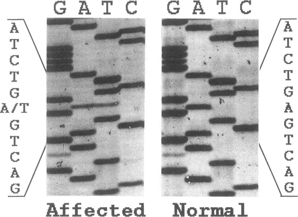
The picture below is copied from the article and it shows the DNA sequencing of this section of the fibrinogen gene for a normal (unaffected) individual on the right and for an affected individual on the left.

Fibrinogen mutation at position 4909

The dark bars indicate which letter is present at each position, corresponding to the letter at the top of that column. For instance, both of these sequences begin at the top with GACTC GGGG. The next letters in the sequence are printed on the far left and the far right. Both sequences continue with ATCTG, but then there is a difference. The Normal pattern on the right has a thick bar in the A column, but the Affected pattern on the left has a thin bar in both the A and the T columns. This indicates one copy of the gene is normal (A) and one copy has substituted a T for an A, hence the "A/T" in the printed column on the far left. (When one copy is normal and one has a mutation, that is called heterozygous. When both copies have the mutation it's called homozygous.)

The four basic building blocks (G, A, T and C) are actually in groups of three which form various amino acids. This A to T substitution changes what is normally GAG (glutamic acid) to GTG (valine) at position 526 of the fibrinogen A alpha chain. That is why this particular fibrinogen mutation is often referred to as Glu526Val.

All seven of the individuals with renal amyloidosis in these two kindreds had this fibrinogen mutation. They also tested 22 individuals who were at risk of having inherited the mutation and found 13 with the mutation and 9 without it.

Since fibrinogen is an important component in the formation of blood clots, they tested clotting times of the affected individuals and found they were all within the normal range. That's good news. They also wanted to determine what percentage of the fibrinogen circulating in the blood was normal fibrinogen and what percentage was mutated fibrinogen. They found it to be essentially 50% normal and 50% mutated.

The Discussion section of the article begins by stating there are several known variants of the fibrinogen A alpha chain that are known to cause dysfibrinogenemia, which is a group of inherited disorders that affect blood clotting in various ways depending on the mutation. (As of 2013 there are over 350 different variants.) Since mutations of the fibrinogen A alpha chain typically affect blood clotting, when you tell you doctors you have a fibrinogen mutation do not be surprised if they ask you if there is any impact on blood clotting. (My primary physician asked me.) Based on the information in this article (and others) you can confidently state that this mutation (Glu526Val) does not affect blood clotting.

The article then discusses the amyloid deposits themselves. In the 1993 article (above) that describes the Arg554Leu mutation, analysis of the fibrinogen in the amyloid deposits showed that only the variant fibrinogen was present in the amyloid deposits. No tissue was available from any individuals in the kindreds in this article, but the authors say it is likely the amyloid deposits due to this mutation also have only variant fibrinogen. This knowledge will be important in understanding how the amyloid deposits are formed.

"The most distinctive feature of amyloidosis in these kindreds is that affected individuals have nephropathy as the major manifestation without evidence of neuropathy." This nephropathy (kidney issues) is in contrast to most types of familial amyloidosis where neuropathy (nervous system issues) is the most common symptom. The article states that only 10 kindreds with hereditary renal amyloidosis have been reported in the literature, and it then gives some basic info on each one, including the mutation if that is known.

The article closes with some comparison between these kindreds and others with hereditary renal amyloidosis. It then states that although both of these kindreds are of Irish descent, a common ancestor could not be found. But it is more likely they have a common ancestor than it is that each family had the mutation occur independently.

=====

So there you have it. If you have the fibrinogen Glu526Val mutation it means the DNA in a reproductive cell of one of your ancestors was copied with a T instead of an A in one specific position. That one minor error was accurately passed along from generation to generation, eventually giving you the potential for kidney problems late in life. Cause and effect can sometimes be a little strange.

The next articles up for review will be some more DNA analysis of other kindreds. We might find which previously reported kindreds with hereditary renal amyloidosis actually ended up having a fibrinogen mutation, and we might find some more fibrinogen mutations as well.

======
Citations for articles discussed in this post:

(1) Benson MD, Liepnieks J, Uemichi T, Wheeler G, Correa R. Hereditary renal amyloidosis associated with a mutant fibrinogen alpha-chain. Nat Genet 1993; 3: 252-255.

(2) Uemichi T, Liepnieks JJ, Benson MD. Hereditary renal amyloidosis with a novel variant fibrinogen. J Clin Invest. 1994;93(2):731-736.


Friday, March 15, 2013

Thrombectomy or Thrombolysis?

It's time to provide an update on Mom due to some very recent activity. In today's episode we'll learn about yet another medical procedure, but first let's start with the beginning of March. The dialysis session on Friday, March 1 went well. They still haven't started the buttonhole technique, but the plan now is to start it later this month after Mom gets back from a trip to Austin and the tech who creates the buttonholes gets back from vacation. It will be nice once that is done since the insertion of the needles is really painful.

Speaking of pain, Mom learned that they can give her a prescription for some Lidocaine cream to apply to her arm before a dialysis session. That will numb the skin a little and thereby reduce the pain when the needles are inserted. It would have been nice to know about that a little sooner.

Toward the end of the dialysis session on Monday, March 4, Mom started having a lot of pain in her arm due to one of the needles. Normally the pain doesn't last once they get them inserted and get dialysis started, but maybe she moved a little to make this one hurt. In any case, the pain was so bad that she called the tech over and said she couldn't continue because she was in so much pain.

On Wednesday, March 6 the tech got Mom started on dialysis and then looked at the machine and said, "Uh oh," which is just what you don't want to hear when your circulatory system is connected to a machine. Mom's blood was going into the machine fine, but it was clogging the machine on the way out. The tech had to clean out the machine, throw a bunch of stuff away, and start over. Fortunately Mom did fine after the restart.

Mom and I did not attend the local amyloidosis support group meeting on Saturday, March 9. She was in Austin and I was traveling to North Carolina.

Since Mom was in Austin she had a couple of dialysis sessions there. The one on Friday, March 8 went great, but on Monday they had an issue with the machine clotting again.

There seem to be a lot of issues with clotting lately, which isn't a total surprise since Mom isn't taking either of the two anticoagulants typically given to patients on dialysis. (That drama unfolded late last year and is covered in the January 30, 2013 post.) So it should come as no surprise that we eventually had a slightly more serious clotting issue.

On Wednesday, March 13, the dialysis machine was clotting frequently and they were having a lot of difficulty with clotting in the blood vessels of Mom's arm as well. They stopped dialysis early and scheduled her to go to Plano Vascular Center the next day for a procedure to clear the clots in her arm and then have dialysis at the center next door. I believe the procedure is either thrombectomy or thrombolysis, which is described in the "How does the procedure work?" section on this page: http://www.radiologyinfo.org/en/info.cfm?pg=dialysisfistulagraft. One procedure uses medication to dissolve the clot and the other one uses a mechanical device.

On Thursday, March 14 my sister Laura took Mom to the appointment at Plano Vascular Center. The doctor there said that she didn't need a thrombectomy or thrombolysis because there was no sign of the fistula clotting. He did, however, perform another balloon angioplasty, this time going a little higher than the most recent one on February 26. So this is the third angioplasty Mom has had on her fistula. They had a little trouble with the needles getting dialysis started, but they got it done and she was fine that night.

On Friday, March 15, she was supposed to have her regularly scheduled Friday dialysis session but they were having so much trouble getting the needles in that she told them to stop and she wouldn't have dialysis today since she had it yesterday. That would mean no dialysis between Thursday and Monday. The nephrologist came by later and said that was not a good idea because her potassium level could get way off going so long without dialysis. Since they are having so much trouble with the fistula he sent her back to Plano Vascular Center where she had another catheter inserted. This one is on her left side instead of right, and it's in her jugular vein so it's higher up. So I guess we're dealing with a permacath once again. Yay.

Here's what the doctor at Plano Vascular Center told Mom and how he recommends she proceed. Since her arm is so badly bruised from all of the attempted needle sticks, he thinks they haven't actually been getting blood just from the fistula but also (or in some cases only) from the bruises (hematomas). That's why they thought her fistula was clotting at the dialysis clinic. He checked her fistula and said it's perfectly fine and actually very strong (good blood flow).

Going forward, he wants her to use just the catheter for dialysis for the next week or two, allowing her fistula and arm to heal. Then hopefully she can have one of the more experienced techs at the dialysis clinic try to access the fistula again. If they still have trouble then she may need to have a procedure where the blood vessels of the fistula are brought closer to the surface of the skin.

We were quite concerned on Wednesday that we would need to have some serious discussions with Mom's hematologist about putting her on some sort of anticoagulant, but now it looks like there may be a different explanation for the clotting issues they are experiencing during her dialysis sessions. Hopefully there will be some good news to report in two to three weeks.

Sunday, March 10, 2013

Article Review (1991) - Inherited Amyloidosis

Today's post reviews an article from 1991. Articles from the previous two decades included a few case reports of what appeared to be a hereditary form of amyloidosis that affects the kidneys, but the search for the underlying genetic cause was still in its infancy. This article was written before the first published findings on a fibrinogen mutation, so the word "fibrinogen" does not appear in this article either.


Author: Dr. Merrill D. Benson (Indiana University School of Medicine, Indianapolis, IN, USA)

Journal: Journal of Medical of Genetics (1991, February)

Here's a link to the PDF of this article, if you'd like to follow along: http://jmg.bmj.com/content/28/2/73.full.pdf+html

The author of this article, Dr. Merrill D. Benson, is very well-known in the amyloidosis community, having discovered the origin of many forms of hereditary amyloidosis. He was one of the doctors at the familial meeting in Chicago in 2011. (He reminds me of Orville Redenbacher.) His lab does genetic testing on a research basis, and it's where one of my sisters sent her blood for testing in May of last year.

This article (no abstract) discusses the various types of hereditary amyloidosis that were known in 1991. It begins with a simple two-sentence definition of amyloidosis that you can memorize to amaze and impress your friends and family. Whenever someone asks you what amyloidosis is you can reply with this: "The amyloidoses are a group of deposition diseases in which the tissue deposits are composed of protein fibrils. These fibrils are the result of aggregation of specific precursor proteins into ordered structures which are resistant to proteolytic digestion and solubilisation." If that's not simple enough for your audience you can use my explanation, which is to think of amyloidosis as teeny tiny pieces of plastic floating around that your body can't get rid of, so they end up collecting in various organs and gumming things up. Ok, back to the article . . .

After a short discussion of localized vs. systemic amyloidosis, the article gives an overview of some different types of hereditary amyloidosis. It starts with familial amyloidotic polyneuropathy (FAP), the most common form of hereditary amyloidosis. FAP is primarily due to a mutation in transthyretin (hence the current designation of ATTR), although at the time this article was published a variant of apolipoprotein A1 had recently been discovered to cause FAP. Then a paragraph is spent listing a few other rare types of hereditary amyloidosis but oddly enough, hereditary renal amyloidosis is not mentioned.

The article then goes into detail on some different types of FAP. They are:

  • Type I: Starts in the lower extremities.
  • Type II: Typically starts with carpal tunnel syndrome and progresses to a more generalized, slowly progressing peripheral neuropathy.
  • Type III: This type of FAP does not present with significant neuropathy early in the course of the disease, but instead will initially affect the heart or the gastrointestinal tract, depending on the mutation.
  • Type IV: A rare Finnish type affecting the cornea and also causing cranial neuropathy.
The article then discusses the various genetic mutations that were known at the time to cause hereditary amyloidosis. As previously mentioned, transthyretin mutations (of which there are many) are the most common cause of hereditary amyloidosis, with the symptoms varying with the mutation. Other mutations covered are apolipoprotein A1, gelsolin, and cystatin C. Again, no mention of fibrinogen since that mutation was yet to be discovered.

The next two sections of the article have the headings Detection of gene carriers and Genetic counselling. This part of the article discusses the impact the current knowledge of molecular genetics has on the study and management of these diseases. I'll list these in bullet form:
  • Allows more in depth studies into the pathogenesis of these diseases (how they develop and progress)
  • Expands the scope of the clinical approach to these diseases
  • Improves diagnosis of these diseases, especially given late onset, incomplete penetrance (not everyone with the genetic mutation will develop symptoms), and opportunities for misdiagnosis due to clinical similarities with other conditions
  • Allows analysis of clinical syndromes among different ethnic groups with identical mutations
  • Genetic testing can be used to determine whether or not someone at risk of having a mutation actually did inherit it.
The final section is Therapy and future prospects. It states up front that there are no specific therapies for any form of hereditary amyloidosis. Preventing the disease at the genetic level would require some mechanism of altering gene expression, but as more is understood about how the fibrils themselves are formed there may be ways of altering the biochemistry to prevent fibril formation in the future. (I believe that is indeed what is happening now with some of the drugs that have been developed for ATTR.) It is particularly important to understand why some subjects develop symptoms so much later than others with the same mutation. (Based on some discussion of that question at the 2011 familial amyloidosis meeting in Chicago, I don't think they are any closer to figuring that out yet.)

The article closes with a paragraph that starts with "although there are no specific therapies for inherited amyloidoses, modern medicine has much to offer to the person afflicted with these diseases." It then lists some medicines and other treatments available for treating peripheral neuropathy, end stage renal disease, and cardiac disease.
So that's the state of familial amyloidosis in 1991, although there is no specific mention of hereditary renal amyloidosis. Perhaps Dr. Benson was considering hereditary renal amyloidosis to be in the category of FAP Type III, which does not present with neuropathy. In any case, we don't have to wait much longer before we see the word "fibrinogen" in an article about amyloidosis.

[Minor update, 11-5-13: typo]
=====

Citation: Benson MD. Inherited amyloidosis. J Med Genet 1991;28: 73-8.

Tuesday, March 5, 2013

Article Reviews - The 1980s

My previous article review actually covered three articles from the 1970s which provided some history of hereditary renal amyloidosis, before the fibrinogen mutation was discovered. This post will cover three more articles, this time from the 1980s. I don't actually have any of these articles, but based on their abstracts and what is said about them in other articles I do have, they seem to have some historical significance.

(Before getting into the articles, I'll note that since this is the first post of the month, the always exciting section on the monthly blog stats will be at the end of this post.)

Since I don't have any of these three articles I'll just give same basic info about each one and then discuss them as a whole.


[Update: The following article was reviewed again on 9-6-13.]


Authors: Mornaghi, Rubinstein, and Franklin

Journal: Transactions of the Association of American Physicians (1981)

I have not found an abstract for this article online, but based on what has been written about it when referred to in other articles I know it is a report on three Irish-American siblings with a clinical history of nephropathy at the ages of 49, 52 and 55.


[Update: The following article was reviewed again on 9-18-13.]


Authors: Lanham, Meltzer, De Beer, Hughes and Pepys (yes, that Pepys)

Journal: QJM: An International Journal of Medicine (1982)

Abstract:
A 23 year old Englishman presented with keratoconjunctivitis sicca and was found to have systemic amyloidosis. Five members of his family in two generations also had non-neuropathic amyloid particularly affecting the kidneys. This conforms to the Ostertag type of hereditary amyloidosis. Amyloid deposits in the proband showed permanganate-sensitive Congophilia and positive immunofluorescence staining for P component, but were negative for amyloid A and prealbumin. These observations suggested that the fibril protein in this patient was immunochemically distinct from the amyloid fibrils characterized hitherto.


[Update: The following article was reviewed again on 9-6-13.]

Title: Familial renal amyloidosis: Case reports and genetic studies (3)

Authors: Mornaghi, Rubinstein, and Franklin

Journal: The American Journal of Medicine (October 1982)

Abstract:
Rapidly progressive biopsy-proved renal amyloidosis developed in three brothers, aged 49, 52, and 55, of Irish-American origin. None had evidence of a plasma cell dyscrasia, a monoclonal serum or urine protein, or any underlying chronic disease. Immunoperoxidase staining of one pulmonary and one renal biopsy specimen was negative for Amyloid A (AA), Amyloid L (AL), and prealbumin. To investigate factors that might play a role in the disease, the subjects and 21 relatives were typed for antigens of the A, B, C, and DR loci and the linked marker genes for factor B and glyoxalase. The ability of macrophages to degrade serum amyloid A (SAA) [1] was examined. One brother yielded an intermediate AA-like produce similar to what is seen in most patients with AA or AL amyloidosis and 40 percent of normal subjects. The other two degraded SAA completely to small peptides. Analysis of the families revealed first that the disease was not linked to the major histocompatibility complex. We were unable to demonstrate a genetic relationship between processing of SAA by peripheral mononuclear cells and the human leukocyte antigen locus. Finally, the pattern of SAA degradation was not associated with the development of the disease.
=====

The two articles authored by Mornaghi are reporting on the same family (Irish-American siblings), so these articles from the 1980s report on two more families with a hereditary form of amyloidosis that primarily affects the kidneys. Most of the medical terminology in the abstracts is beyond my understanding, but here are some key points I picked up:


  • The abstract of the Lanham article indicates they determined the amyloid fibrils themselves are different than amyloid fibrils previously analyzed. I assume the previously analyzed fibrils were from patients with the most common form of amyloidosis, known as primary or AL amyloidosis.
  • The Mornaghi report indicates they also determined these patients did not have other types of amyloidosis that were known at the time, specifically AL and AA amyloidosis. This report also appears to describe a significant effort to determine the genetics behind this form of amyloidosis by doing various types of analysis on 21 relatives of these three siblings.

So that was the state of things in the early 1980s. A few isolated families in the US and England have been determined to have a hereditary form of amyloidosis that affects the kidneys, and the amyloid fibrils (the "stuff" the damages the kidneys) appear to be different than the fibrils associated with other more common types of amyloidosis. The symptoms and progression of the disease are being documented, as are the detailed pathological findings of analyzed tissue samples.

In really simple terms, the medical community at this point knows "what" is going on but they don't know "why." The cause is likely genetic, but they are a long way from determining the specific genetic cause (or causes). We'll need a few more years for the knowledge of genetics and the available technology to advance so medical researchers can zero in on the genetic cause. In fact, the next article I'll be reviewing is "Inherited Amyloidosis" from 1991.

=====Monthly Blog Status Update
Months behind as of February 2013: 2
Months behind as of March 2013: zero!

The "Months Behind" blog stat can officially be retired now that I have caught up to the present day. I'm not sure what to replace it with, so for now I'll go with something easy, which is pageviews.


Total posts: 64 (8 in February)

Total pageviews: 1016 (~120 in February)


Email subscribers: 4 (Don't worry. I can't see the email addresses used to subscribe, only the count.)


Number of countries that have viewed the blog: 26


New countries that viewed the blog in February:

Taiwan
Spain
New Zealand
==========

Citations for the articles discussed in this post:

(1) Mornaghi, R., P. Rubinstein, and E. C. Franklin. 1981. Studies on the pathogenesis of a familial form of renal amyloidosis. Trans. Assoc. Am. Phys. 94:21 1-216.

(2) Lanham, J. G., M. L. Meltzer, F. C. De Beer, G. R. V. Hughes, and M. B. Pepys. 1982. Familial amyloidosis of Ostertag. Q. J. Med. 201:25-32.

(3) Mornaghi, R., P. Rubinstein, and E. C. Franklin. 1982. Familial renal amyloidosis: case reports and genetic studies. Am. J. Med. 73:609-614.